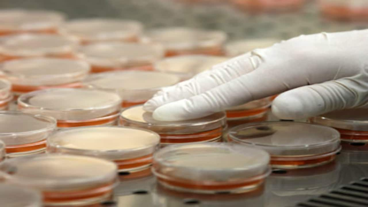
Carrier
tests:
These
tests are the oldest tests. The test described by Robert Koch was a carrier
test. In these tests, the carrier such as a silk or catgut thread or a
penicylinder (a little stick) is contaminated by submersion in a liquid culture
of the test organism. The carrier is then dried and is brought in contact with
the disinfectant for a given exposure time. After the exposure, it is cultured
in a nutrient broth; no growth indicates activity of the disinfectant tested
whereas growth indicates a failing. By multiplying the number of test
concentrations of the disinfectant and the contact times, a potentially active
concentration-time relationships of the disinfectant is obtained. Example of a
carrier test is the former use-dilution test of the American Association of
Official Analytical Chemists (AOAC, 1990). Limitation of the carrier tests are:
a) the number of bacteria dried on a carrier is hard to standardize and b) the
survival of the bacteria on the carrier during drying is not constant. The AOAC
Use-dilution test is a carrier-based test. The organisms used are Salmonella
cholerasuis, S. aureus and P. aeruginosa. Carriers (stainless steel cylinders)
are meticulously cleaned, sterilized by autoclaving in a solution of aspargine,
cooled and inoculated with a test organism by immersing in one of the culture
suspensions. The cylinders are drained on filter paper, dried at 37o C for 40
minutes, exposed to the use-dilution of the disinfectant for 10 minutes, and
cultured to assess the survival of the bacteria. A single test involves the
evaluation of 60 inoculated carriers (one organism) against one product sample.
In addition to the 60 carriers, 6 carriers are required to estimate carrier
bacterial load and 6 more are included as extras. Thus, a total of 72 seeded
carriers are required to perform a single test. A result showing no growth in all
ten tubes confirms the result of phenol coefficient test. If any carrier
produces growth, the test must be repeated using a lower dilution of the
disinfectant. Use-dilution test is performed to confirm the efficiency of
disinfectant dilution derived from phenol coefficient test.
Suspension tests:
In these tests, a sample of the bacterial culture
is suspended into the disinfectant solution and after exposure it is verified
by subculture whether this inoculum is killed or not. Suspension tests are
preferred to carrier tests as the bacteria are uniformly exposed to the
disinfectant. There are different kinds of suspension tests: the qualitative
suspension tests, the test for the determination of the phenol coefficient
(Rideal and Walker, 1903) and the quantitative suspension tests. Initially this
was done in a qualitative way. A loopful of bacterial suspension was brought
into contact with the disinfectant and again a loopful of this mixture was
cultured for surviving organisms. Results were expressed as ‘growth’ or ‘no
growth’. In quantitative methods, the number of surviving organisms is counted
and compared to the original inoculum size. By subtracting the logarithm of the
former from the logarithm of the latter, the decimal log reduction or
microbicidal effect (ME) is obtained. An ME of 1 equals to a killing of 90% of
the initial number of bacteria, an ME of 2 means 99% killed. A generally
accepted requirement is an ME that equals or is greater than 5: at least
99.999% of the germs are killed. Even though these tests are generally well
standardized, their approach is less practical.
Determination of phenol
coefficient:
Phenol coefficient of a disinfectant is calculated
by dividing the dilution of test disinfectant by the dilution of phenol that
disinfects under predetermined conditions.
Rideal Walker method:
Phenol is diluted from 1:400 to
1:800 and the test disinfectant is diluted from 1:95 to 1:115. Their
bactericidal activity is determined against Salmonella typhi suspension.
Subcultures are performed from both the test and phenol at intervals of 2.5, 5,
7.5 and 10 minutes. The plates are incubated for 48-72 hours at 37°C. That
dilution of disinfectant which disinfects the suspension in a given time is
divided by that dilution of phenol which disinfects the suspension in same time
gives its phenol coefficient.
For example, after 7.5 minutes, the test organism
was killed by the test disinfectant at a dilution of 1;600. In the same period
the test organism was killed by phenol at a dilution of 1:100.
Phenol coefficient = 600/100 = 6
This result indicates
that the test disinfectant can be diluted six times as much as phenol and still
possess equivalent killing power for the test organism. Disadvantages of the
Rideal-Walker test are: No organic matter is included; the microorganism
Salmonella typhi may not be appropriate; the time allowed for disinfection is
short; it should be used to evaluate phenolic type disinfectants only.
Chick Martin test:
This test also determines the phenol coefficient of
the test disinfectant. Unlike in Rideal Walker method where the test is carried
out in water, the disinfectants are made to act in the presence of yeast
suspension (or 3% dried human feces) to simulate the presence or organic
matter. Time for subculture is fixed at 30 minutes and the organism used to test
efficacy is S.typhi as well as S.aureus. The phenol coefficient is lower than
that given by Rideal Walker method.

The phenol coefficient test recommended by AOAC
included two test organisms (S. aureus and P. aeruginosa) and included the
disinfectant inactivators in the recovery medium. The recovery medium Letheen
broth contains the inactivator Lecithin and Polysorbate 80. In separate tests,
the bacterial suspensions are added to standard dilutions of pure
phenol and several dilutions of the test disinfectant. After contact time of 5,
10 and 15 minutes, samples are transferred to the recovery medium by a standard
wire loop. When the positive and negative cultures have been recorded, the
result of the test is expressed as phenol coefficient. It is calculated by
dividing the highest dilution of the disinfectant that kills the test inoculum
in ten minutes but not in five minutes by the dilution of phenol that gives the
same result.
Disinfectant kill time test
This test was designed to demonstrate log reduction
values over time for a disinfectant against selected bacteria, fungi, and/or
mold. The most common organisms tested include: Bacillus subtilis, Bacillus
atrophaeus, Bacillus thuringiensis, Staphylococcus aureus, Salmonella
cholerasuis, Pseudomonas aeruginosa, Aspergillus niger, and Trichophyton
mentagrophytes. A tube of disinfectant is placed into a waterbath for
temperature control and allowed to equilibrate. Once the tube has reached
temperature, it is inoculated to achieve a concentration of approximately 106
CFU/mL. At selected time points (generally five points are used including zero)
aliquots are removed and placed into a neutralizer blank. Dilutions of the
neutralizer are made and selected dilutions plated onto agar. Colonies are
enumerated and log reductions are calculated.
Capacity tests:
Each time a soiled instrument is placed into a
container with disinfectant, a certain quantity of dirt and bacteria is added
to the solution. The ability to retain activity in the presence of an
increasing load is the capacity of the disinfectant. In a capacity test, the
disinfectant is challenged repeatedly by successive additions of bacterial
suspension until its capacity to kill has been exhausted. Capacity tests
simulate the practical situations of housekeeping and instrument disinfection.
The best known capacity test is the Kelsey-Sykes test (Kelsey and Sykes, 1969).
Kelsey-Sykes test is a triple challenge
test, designed to determine concentrations of disinfectant that will be
effective in clean and dirty conditions. The disinfectant is challenged by
three successive additions of a bacterial suspension during the course of the
test. The duration of test takes over 30 minutes to perform. The concentration
of the disinfectant is reduced by half by the addition of organic matter
(autoclaved yeast cells), which builds up to a final concentration of 0.5%.
Depending on the type of disinfectant, a single test organism is selected from
S. aureus, P. aeruginosa, P. vulgaris and E. coli. The method can be carried out
under 'clean' or 'dirty' conditions. The dilutions of the disinfectant are made
in hard water for clean conditions and in yeast suspension for dirty
conditions. Test organism alone or with yeast is added at 0, 10 and 20 minutes
interval. The contact time of disinfectant and test organism is 8 min. The
three sets of five replicate cultures corresponding to each challenge are
incubated at 32o C for 48 hours and growth is assessed by turbidity. The
disinfectant is evaluated on its ability to kill microorganisms or lack of it
and the result is reported as a pass or a fail and not as a coefficient. Sets
that contain two or more negative cultures are recorded as a negative result.
The disinfectant passes at the dilution tested if negative results are obtained
after the first and second challenges. The third challenge is not included in
the pass/fail criterion but positive cultures serve as inbuilt controls. If
there are no positive cultures after the third challenge, a lower concentration
of the disinfectant may be tested.


The capacity test of Kelsey and Sykes gives a good
guideline for the dilution of the preparation to be used. Disadvantage of this
test is the fact that it is rather complicated.
Test for stability and long-term effectiveness:
Recommended concentrations based on Kelsey Sykes
test apply only to freshly prepared solutions but if the solutions are likely
to be kept for more than 24 hours, the effectiveness of these concentrations
must be confirmed by a supplementary test for stability of unused solution and
for the ability of freshly prepared and stale solutions to prevent
multiplication of a small number of bacteria that may have survived the short
term exposure. P. aeruginosa is used a test organism. Sufficient disinfectant
solution is prepared for two tests. One portion is inoculated immediately and
tested for growth after holding for seven days at room temperature. The other
portion is kept at room temperature for seven days and then inoculated with a
freshly prepared suspension of test organism. It is also tested for growth
seven days after inoculation. If growth is detected, a higher concentration of
disinfectant must be tested in the same way.
Practical tests:
The practical tests under real-life conditions are
performed after measuring the time-concentration relationship of the
disinfectant in a quantitative suspension test. The objective is to verify
whether the proposed use dilution is still adequate in the conditions under
which it would be used. The best known practical tests are the surface
disinfection tests. Surface tests assess the effectiveness of the selected
sanitizer against surface-adhered microorganisms. The test surface (a small
tile, a microscopic slide, a piece of PVC, a stainless steel disc, etc.) is
contaminated with a standardized inoculum of the test bacteria and dried: then
a definite volume of the disinfectant solution is distributed over the carrier;
after the given exposure time the number of survivors is determined by
impression on a contact plate or by a rinsing technique, in which the carrier
is rinsed in a diluent, and the number of bacteria is determined in the rinsing
fluid. In order to determine the spontaneous dying rate of the organisms caused
by drying on the carrier, a control series is included in which the
disinfectant is substituted by distilled water; from the comparison of the
survivors in this control series with the test series, the reduction is
determined quantitatively.
There is an essential
difference between a carrier test and a surface disinfectant test: in the
former case the carrier is submerged in the disinfectant solution during the
whole exposure time, whereas in the latter case the disinfectant is applied on
the carrier for the application time and thereafter the carrier continues to
dry during the exposure. Surface tests can reflect in-use conditions like
contact times, temperatures, use-dilutions, and surface properties.
Surface Time kill Test
A 24 hour culture in nutrient broth culture is
prepared. A volume of microbial culture (usually 0.010 mL to 0.020 mL) is
placed onto the center of each of a number of sterile test surfaces. This
inoculum can be spread over the sterile test surface in a circular pattern to
achieve a thin, uniform coverage with the test microorganism if desired. To measure
initial microbial concentrations, one or more untreated, inoculated test
surfaces are harvested and microorganisms are enumerated. The remaining
inoculated test surfaces are treated with the disinfectant, each for a
different length of time. Immediately after the treatment times have elapsed,
the test surfaces are placed into a solution that neutralizes the disinfecting
action of the product, and microorganisms surviving treatment with the
disinfectant or sanitizer are cultured and enumerated. Results of the timekill
study are tabulated and reported, usually by charting microbial concentrations
on the test surfaces as a function of treatment time with the disinfectant or
sanitizer.
In-use test:
A simple to use test was described by Maurer in
1985 that can be used in hospitals and laboratories to detect contamination of
disinfectants. A 1 ml sample of the disinfectant is added to 9 ml diluent which
also contains an inactivator. Ten drops, each of 0.02 ml volume of the diluted
sample are placed on each of two nutrient agar plates. One is incubated at 37o
C for three days and the other at room temperature for seven days. Five or more
colonies on either plate indicate contamination.
British standard tests for quaternary ammonium
compounds:
This test was initially described in 1960 to
distinguish bactericidal action from the high level of bacteriostatic activity,
which is characteristic of QACs. This test is also applicable to other
bactericides such as chlorhexidine and synthetic phenols. The inactivator used
contains 2% lecithin and 3% non-ionic detergent (polysorbate 80). The test is
performed using suspensions of gram negative and gram positive bacteria with or
without the inclusion of organic matter. If a series of samples are taken from
a dilution of the disinfectant containing 5 x10⁸ to 5 x 10⁹ bacteria per ml at
the start of the test, a death curve may be prepared from the colony counts on
the agar containing recovery medium and reduction factors up to 106 (99.99%
kill) can be verified. In order to determine the antimicrobial value of QAC, it
was revised as the highest dilution of the disinfectant that, under the test
conditions, will reduce the microbial population to a colony count not greater
than 0.01% of that in the control. In this revision, E. coli was used and the
contact time was 10 minutes. The challenge medium is E. coli culture suspension
with equal amount of horse serum. One ml of the challenge is added to nine ml
of each dilution of the disinfectant along with two control tubes containing diluent
alone. At the end of exposure period, one ml each of the mixture is added to 9
ml of inactivator and the surviving bacteria are counted as colony forming
units on agar plates.
Testing schemes:
The antimicrobial efficiency of a disinfectant is
examined at three stages of testing. The first phase concerns laboratory tests
in which it is verified whether a chemical compound or a preparation possesses
antimicrobial activity: for these preliminary screening tests essentially
quantitative suspension tests are considered. The second stage is still carried
out in the laboratory but in conditions simulating real-life conditions. Not
disinfectants, but disinfection procedures are examined. It is determined in
the practical tests in which conditions and at which use-dilution after a given
contact time the preparation is active. The third phase comprises the field
tests or pilot studies, and the variant of in-use tests. In these tests it is
verified whether, after a normal period of use, germs in the disinfectant
solution are still killed. Most studied are the bactericidal tests in which the
activity towards vegetative bacteria is examined. AOAC has schedules that are
applicable for fungi and yeasts too (fungicidal tests), for mycobacteria
(tuberculocidal tests), for viruses (virucidal tests) and for spores of
bacteria (sporicidal tests).
Bactericidal tests:
A bactericidal test must include the following
sequence of steps: 1. The test organism is exposed to a suitable concentration
of the disinfectant 2. Samples are taken at specified times and added
immediately to a diluent or culture medium containing the appropriate
disinfectant inactivator 3. The treated samples are cultured for surviving
microorganisms.
Test organisms:
Specified strains (usually ATCC) of S. aureus, P.
aeruginosa, P. vulgaris and E. coli are usually recommended. A synthetic broth
is recommended for preparing a series of subcultures to be used in the tests.
The 24-hour broth culture may be used without further treatment; however, it is
usually filtered (to remove slime) and centrifuged. The washed bacteria are
resuspended in hard water to which autoclaved yeast or serum may be added to
simulate dirty conditions. Finally, the suspension is shaken with glass beads
on a vortex mixer and a viable count is set up immediately before performing
the test.
The disinfectant:
The concentration or dilution of the disinfectant
to be tested may be based on manufacturer’s recommendations. The solutions
should be prepared on the day of test. Distilled water or standard hard water
is used to make dilutions. Tap water is unsuitable because it may contain
chemicals that may precipitate with some disinfectants.
References:
The
testing of disinfectants: Gerald Reybrouck, International Biodeterioration
& Biodegradation 41 (1998) 269-272
Introduction
to sterilization and disinfection control, 2nd edition, Churchill Livingstone Joan
F.Gardner, Margaret M Peel. 1991